Step 3 of 3. Imagine an action that you engage in every day and explain how neurons and neurotransmitters might work.

12 2 Nervous Tissue Anatomy Physiology
Fasicles - a bundle of structures such as nerve or muscle fibers.
. Covers the choroid plexus. Draw a typical multipolar neuron in the space below. Cell body nucleus nucleolus chromatophilic substance dendrites axon myelin sheath myelin sheath gaps and axon terminals.
View a sample solution. Include and label the following structures on your diagram. A neuron is composed of an axon dendrites and soma cell body.
Up to 24 cash back 1. WHAT SUBSTANCE IS FOUND IN SYNAPTIC VESICLES OF. Neurons are specialized cells that transmit electrical signals in the brain.
E А B С D RNA transcription receives excitatory or inhibitory input only dendrites contains voltage gated ion channels for action potentials axon terminal contains. The neuron master shi heng. Match the structures and functions listed below with the 5 regions or combinations of regions.
Draw a typical Neuron and label the following. Draw a typical neuron in the space below. View this answer View this answer View this answer done loading.
Slightly rounded co amp 8230 draw a typical multipolar neuron in the space below include and label the following structures on your diagram cell body nucleus nucleolus nissl bodies dendrites axon axon collateral branch myelin sheath nodes of. Start studying multipolar neuron. Any voltage change in that direction makes a neuron more likely to fire.
Question 1 6 points The multipolar neuron below has been divided into 5 regions labelled A-E. Neuron draw a picture of a typical neuron and label its parts draw the human cell with the label pdf free download here cell structure bemidji state draw a typical multipolar neuron in the space below include and label the following structures cell body nucleus dendrites axon myelin sheath the human brain related ebooks the cell body soma is the factory of the neuron it produces all. Label the parts of a typical multipolar neuron.
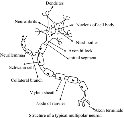
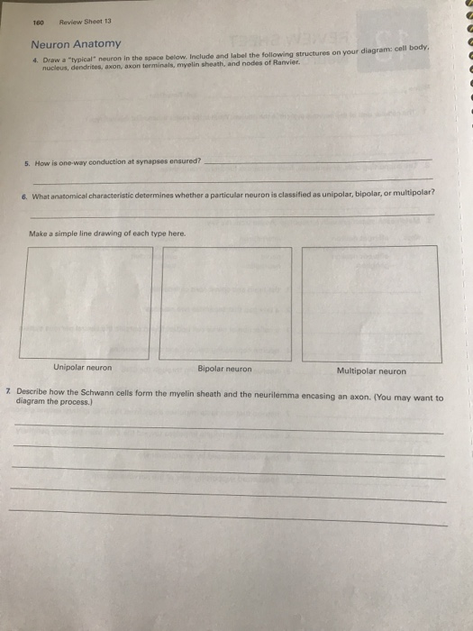
Lines space in the brain ventricles Ependymal cells. Cell body soma Dendrites Axon Nissl bodies Nucleus Nucleolus Axon of Hillock Axonal terminal synaptic knobs Draw the direction in which impulses flow 2 What anatomical characteristic determines whether a particular neuron is classified as unipolar bipolar or multipolar. February 28th 2018 - Draw a typical multipolar neuron draw a typical neuron in the space belowraw a typical ne Its how Clinton has all ready to vote for Hillary and thenstreaming missioncollege org May 6th 2018 - Review Sheet 13 160 Neuron Anatomy 4 Draw a typical neuron in the space below.
Match the description with the correct classification of neuron. Draw a typical multipolar neuron in the space below. The primary components of the neuron are the soma cell body the axon a long slender projection that conducts electrical impulses away from the cell body dendrites tree-like structures that receive messages from other neurons and synapses.
Cell body nucleus dendrites axon axon terminals myelin sheath and nodes of. Carry impulses from the CNS to effectors. - the space between neurons at a nerve synapse across which a nerve impulse is transmitted by a neurotransmitter.
Cell body nucleus dendrites axon axon terminals myelin sheath and nodes of Ranvier. Neuroglia non-neuronal cells assists the nerve impulses propagation and also provides nutrients to the nerve cells. Axon collateral - side branch of the axon.
DRAW A TYPICAL MULTIPOLAR NEURON. Currently I am unfolding before you twelve simple 3D nail artwork designs ideas trends stickers. Multipolar Neuron Unipolar Neuron Bipolar Neuron.
What anatomical characteristic determines whether a particular neuron is. How is one-way conduction at synapses ensured. They are the functional unit of the nervous system.
Draw a typical neuron in the space below. Draw a picture of a neuron and label its main parts. Chapter 15 Problem 5E is solved.
Draw a typical multipolar neuron in the space below New and most recent designs are now being released by specialists so Increasingly more girls can Keep to the streak of nail art. Include and label the following structures on your diagram. Include and label the following structures on your diagram.
Three-liter flask and boiled. Multipolar neuron in the space below include and label the following structures on your diagram cell body nucleus nucleolus nissl bodies dendrites axon axon collateral branch myelin sheath nodes of ranvier axon terminals and. Learn vocabulary terms and more with flashcards games and other study tools.
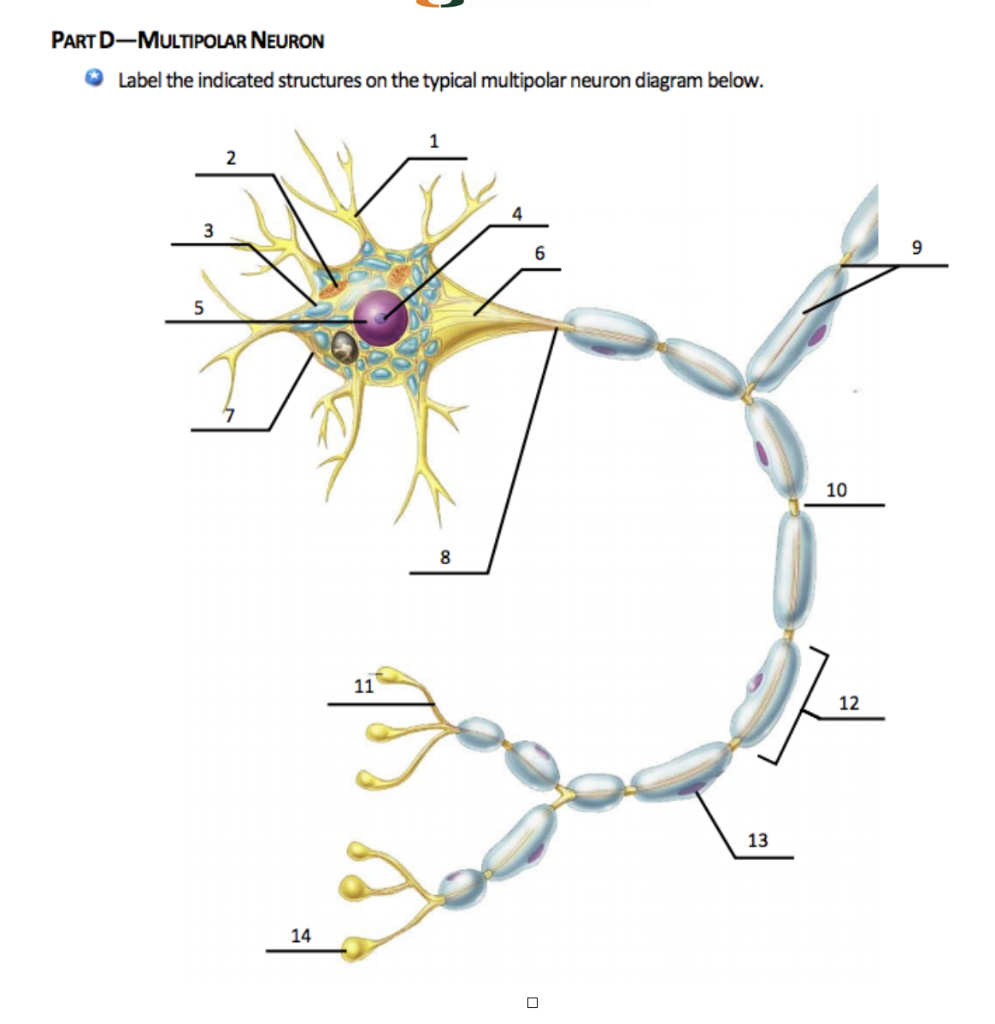
CELL BODY NUCLEUS NUCLEOLUS nISSL bODIES DENDRITES AXON AXON COLLATERAL BRANCH MYELIN SHEATH NODES OF RANVIER AXON TERMINALS AND NEUROFIBRILS. Step 2 of 3. INCLUDE ANDD LABEL THE FOLLOWING STRUCTURES ON YOUR DIAGRAM.
Cell body nucleus nucleolus Nissl bodies dendrites axon axon collateral branch myelin sheath nodes of Ranvier axon terminals and neurofibrils. Draw A Typical Multipolar Neuron In The Space Below. Solved Draw A typical Multipolar Neuron In The Space November 20th 2020 - Draw A typical Multipolar Neuron In The Space Below Include And Label The Following Structures On Your Diagram Cell Body Nucleus Nucleolus Nissl Bodies Dendrites Axon Axon Collateral Branch Myelin Sheath Nodes Of Ranvier Axon Terminals And Neurofibrils.
Usually results from Na flowing into the cell and neutralizing some of the negative charge on the inside of the membrane. Include and label the following structures on your diagram.

Biol 2022 Nervous System Flashcards Quizlet
Chapter 7 Solutions Human Physiology 15th Edition Chegg Com

Ch 17 Histology Of Nervous Tissue Flashcards Quizlet

Nervous Tissue Anatomy And Physiology I
Solved 160 Review Sheet 13 Neuron Anatomy 4 Draw A Typical Chegg Com
Solved Draw A Typical Multipolar Neuron In The Space Below Include And Course Hero

Typical Multipolar Neuron Download Scientific Diagram
Solved Part D Multipolar Neuron Label The Indicated Chegg Com
0 comments
Post a Comment